The Concurrence of Multiple Sclerosis and Glioma
Abstract
The concurrence of multiple sclerosis (MS) and glioma is rare, although it must be kept in mind when an atypical lesion develops in a patient with MS. Indeed, pseudo-tumoral MS lesions may resemble gliomas, and conversely early-stage gliomas may mimic MS. Their entangled presentation may thus pose a significant diagnostic and therapeutic challenge that must be addressed early to enable the best patient outcome.
We here present two cases followed by a literature review:
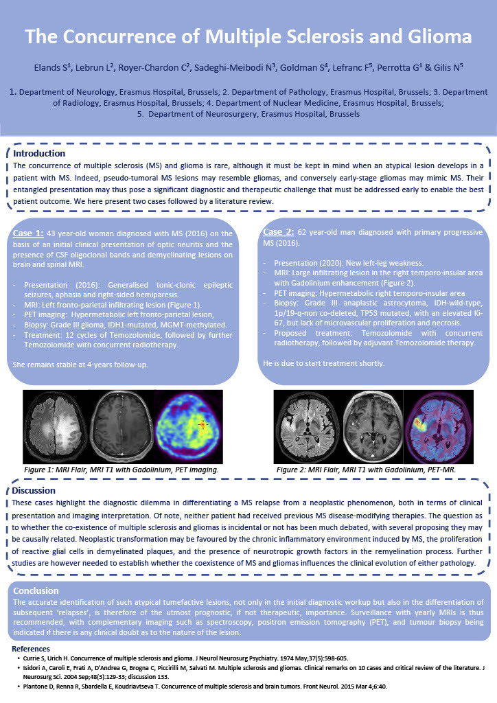
A 43 year-old woman with a diagnosis of multiple sclerosis since one year presented with generalised tonic-clonic epileptic seizures, right-sided hemiparesis and aphasia. MRI identified a left parietal infiltrating lesion, hypermetabolic on PET imaging. Biopsy revealed a grade III glioma, IDH1-mutated, MGMT-methylated. She was treated with temozolomide and concurrent radiotherapy, and remains stable at 4-years follow-up.
A 62 year-old man diagnosed with progressive multiple sclerosis since four years presented with a new left-leg weakness. MRI revealed a large infiltrating lesion in the right temporo-insular area, hypermetabolic on PET imaging, with histopathology confirming a grade IV glioma, IDH-wildtype, 1p/19-q-non co-deleted and TP53 mutated. He is due to start treatment shortly.
The question as to whether the coexistence is incidental or not has been much debated, with several proposing they may be causally related. Neoplastic transformation may be favoured by the chronic inflammatory environment induced by MS, the proliferation of reactive glial cells in demyelinated plaques, and the presence of neurotropic growth factors in the remyelination process. Further studies are needed to establish whether the coexistence of MS and gliomas influences the clinical evolution of either pathology, although immunosuppressive therapy given for the neoplasm tends to reduce disease activity in MS. The accurate identification of such atypical tumefactive lesions, including the role of PET imaging, is therefore of the utmost prognostic, if not therapeutic, importance.